To support the abdominal and pelvic viscera.
Pelvic floor anatomy ct.
Learn the diagnosis of ct and methods of computed tomography.
Gross anatomy the perineum is bounded by the pubis anteriorly the ischial tuberosities anterolaterally the sacrotuberous ligaments posterolaterally and the coccyx posteriorly.
This anatomical chart beautifully illustrates and outlines the nuances of subjects including.
The pelvic floor is a dome shaped muscular sheet separating the pelvic cavity above from the perineal region below.
The male pelvis is different from a female s.
Mdct of the abdomen and pelvis.
15 liver 16 oesophagus 17 stomach 41 descending abdominal aorta 43 inferior vena cava 55 thoracic vertebra.
This cavity encloses the pelvic viscera bladder intestines and uterus in females.
It attaches to the walls of the lesser pelvis separating the pelvic cavity from the perineum inferiorly region which includes the genitalia and anus.
The main function of the pelvic floor muscles are.
We created an anatomical atlas of abdominal and pelvic ct which is an interactive tool for studying the conventional anatomy of the normal structures based on a multidetector computed tomography.
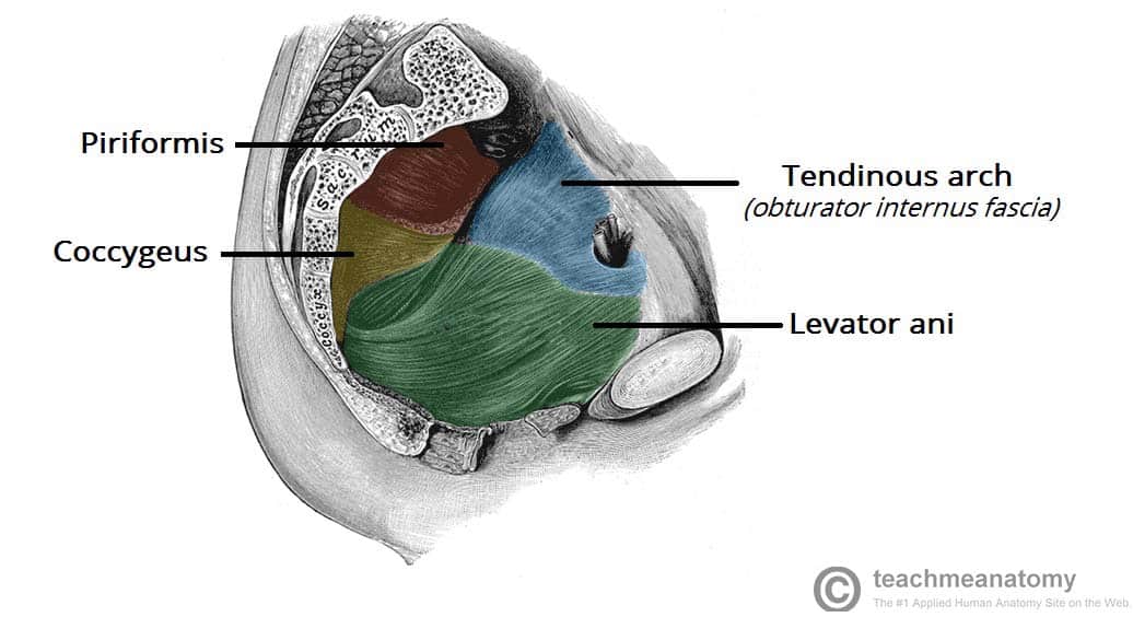
The levator ani muscle also known as the muscular pelvic diaphragm is the musculotendinous sheet that forms the majority of the pelvic floor supports the pelvic viscera and aids in urinary and fecal evacuation as well as maintaining continence.
Anatomical structures of the abdomen and pelvis are visible as interactive labeled images.
The pelvic floor is a funnel shaped structure.
The pelvic region is the area between the trunk or main body and the lower extremities or legs.
The spatial relationship of the organs and the pelvic floor are important.
The lower end of the pelvic floor is held closed by the pelvic floor muscles preventing prolapse by constricting the base.
The pelvic floor musculature anatomy chart shows from multiple angles the way in which the pelvic floor muscles are layered in your body and how they operate in conjunction with adjacent organs from the urinary system reproductive system and more.
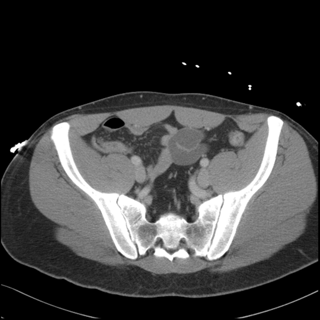
Anatomy ct axial abdomen and pelvis male male abdomen and pelvis ct scan form no 1.
There are two holes that have significance.
Pelvic support is a combination of constriction suspension and structural geometry.