Suggested protocol for dynamic mri of pelvic floor dysfunction.
Pelvic floor mri protocol.
7 high temporal resolution and excellent contrast make it well suited for evaluation of organ movement.
Please have the patient void their bladder prior to exam to improve image quality.
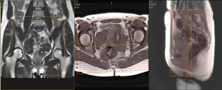
Angle the position block parallel to the lumbar spine.
Slices must be sufficient to cover the para aortic area from mid abdomen to the sacrum.
The patient is asked to defecate while on the mr scanner table and then asked to go to the toilet to completely empty the urinary bladder rectum or rectocele.
For dynamic mri of the pelvic floor use steady state imaging sequences.
Imaging is optimally performed after 3 hours of fasting to reduce bowel peristalsis and following administration of an antiperistaltic agent unless contraindicated.
Diagnostic and biopsies breast imaging protocols currently applied in our mri section.
Plan the big fov coronal slices on the sagittal plane.
For all pelvic mri studies except the bladder protocol or the mr urogram.
Dynamic pelvic floor mri imaging technique the mr imaging evaluation is performed with the patient in the supine position without contrast agents and within fifteen minutes.
A pelvic mri scan specifically helps your doctor to see the bones organs blood vessels and other tissues in your pelvic region the area between your hips that holds your reproductive organs and.
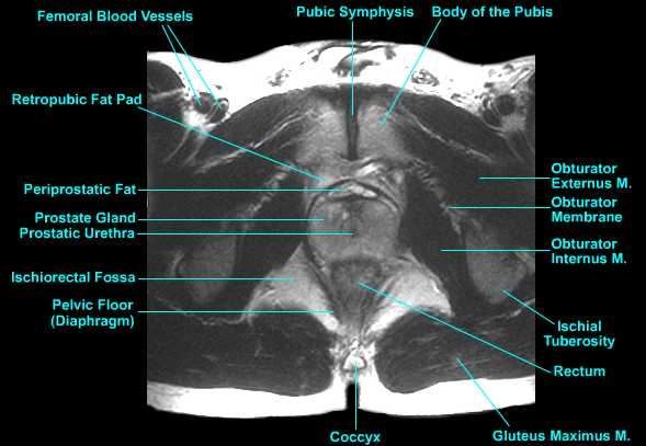
Dynamic pelvic mr allows radiologists to directly see detailed images of the anatomy of the pelvic floor structures which allows analysis of anatomy and function.
Dynamic pelvic floor magnetic resonance imaging mri is a noninvasive test that uses a powerful magnetic field radio waves and a computer to produce detailed pictures of the pelvic floor a network of muscles that stretches between the pubic bone and spine and the abdominal organs it supports.
Endometrial carcinoma dr mostafa el feky and dr laura fender et al.
Check the positioning block in the other two planes.
Studies have shown that dynamic pelvic mri is more sensitive than physical examination making it the gold standard for diagnosing pelvic floor disorders.
Interpretation of mri findings the level of the pelvic floor on dynamic mri can be demarcated radiologically on the midsagittal image using the pubococcygeal line as described by yang et al.
Functional disorders of the pelvic floor such as pelvic organ prolapse and defecatory dysfunction represent a common health problem especially in women it is estimated that more than 15 of multiparous women 1 are affected by some sort of pelvic disorder and that 10 20 of patients seek medical care in gastrointestinal clinics for evacuation dysfunction 2.